Microscopy - Equipments
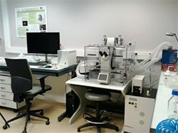
SP8 Confocal Microscope (Leica)
The system in reverse configuration is equipped with 4 lasers covering excitation wavelengths 405, 488, 561, and 638nm. Fluorescence signals are collected on 2 PMT detectors and 2 high-sensitivity HyD detectors. A thermostatic and CO2-regulated chamber and a motorized turntable are installed on the microscope for kinetic studies and XYZT acquisitions.
This microscope is suitable for confocal, wide-field and/or multi-position imaging.
Objectives: HC PL FLUOTAR 5x/0.15 HC PL APO 10x/0.40 CS HC PL APO 20x/0.75 CS2HC Fluotar L 25x/0.95W VISIR HC PL APO 40x/1.30 Oil CS2 HC PL APO 63x/1.40 OIL CS2
Confocal SPE Microscope (Leica)
The system in reverse configuration is equipped with 4 lasers in the "visible" range covering excitation wavelengths 405, 488, 533, and 635nm. Fluorescence signals are collected between 410nm and 850nm. The instrument with a single detector works by sequenced acquisitions. The microscope is equipped with a thermostatic and CO2-regulated chamber for real-time kinetic studies and 4D acquisitions.
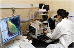
Multiphoton Microscope SP8 (Leica)
Specifically dedicated to the imaging of living organisms and the imaging of thick samples, this inverted system offers a mixed confocal and multiphoton configuration. It is equipped with 2 spectral internal detectors, 2 high-sensitivity internal hybrid detectors, and 4 non-descaned external detectors. In confocal configuration the illumination is carried out with 4 solid lasers (405, 488, 552 and 638nm) and in multiphoton version with the Mai-Tai HP DS tunable laser from 690 to 1040 nm (Spectra Physics). The instrument also includes a resonant scanner for high-speed acquisitions, a motorized stage for wide field and/or multi-position imaging, and a 3D Viewer.
Objectives: HC PL FLUOTAR 5x/0.15 HC PL APO 10x/0.40 CS HC PL APO 20x/0.75 CS2HC Fluotar L 25x/0.95 W VISIR HC PL APO 40x/1.30 Oil CS2 HC PL APO 63x/1.40 OIL CS2
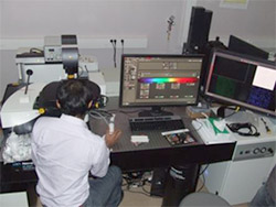
Spinning disk Microscope
This inverted microscope equipped with a Yokogawa-CSU W1 spinning head, is perfectly adapted to long-lasting kinetics. Equipped with a thermo-regulated chamber, CO2 controlled and a motorized stage, it offers the optimal conditions for cellular and sub-cellular dynamic imaging. Laser excitation sources cover the spectrum from UV to far red (405nm, 488nm, 561nm, 642nm). Image acquisition is carried out via 1 Zyla sCMOS camera (82% quantum efficiency, pixel 6.45x6.45 μm). This system is controlled by the Metamorph software, offering many possibilities for acquisition configuration.

Spinning disk Microscope - Live SR
This inverted microscope, equipped with a Yokogawa-CSU W1 spinning head, is perfectly suited for long-lasting kinetics. Equipped with a thermo-regulated and controlled CO2 chamber and a motorized stage, it offers optimal conditions for cellular and sub-cellular dynamic imaging. Laser excitation sources cover the spectrum from UV to far red (405nm, 445nm, 488nm, 515nm, 561nm, 642nm). Image acquisition is carried out via 2 cameras: Orca Flash4 sCMOS (85% quantum efficiency, pixel size 6.45x6.45 μm) and Prime 95B sCMOS (95% quantum efficiency, pixel 11x11 μm). The system is equipped with a super resolution module (Live-SR) that improves the optical resolution by a factor of 1.7. TIRF, FRAP and FRET options can also be combined.
The iLas Modular system allows complete control of the illumination laser source and combines: TIRF 360°; FRAP; photo-activation and photo-ablation. The microscope is controlled by Metamorph 7 software, offering many possibilities for acquisition configuration.
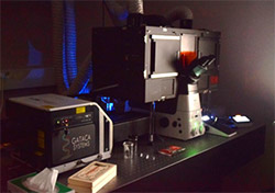
Wide-field microscope (Olympus/Microvision)
This microscope in fully motorized straight configuration (Provis Olympus) is dedicated to the mapping of colored histological sections or any sample marked by fluorophores. Single field acquisition in image stacks and/or multicolors is also possible. The system is equipped with a motorized stage and 7 lenses (from 4x to 100x) as well as two cameras (Sony XCD-U100CR color camera, and Hamamatsu ORCA-ER B&W camera). The image acquisition and processing software Histolab and Archimed, (Microvision) allow carrying out in real time, at acquisition, various measurements on tissues or cells: median, surface, coloration density, counting and the corresponding statistical analyses.
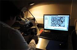
AZ100 Fluorescence Macroscope (Nikon)
Very flexible, this macroscope allows fluorescence and trans-illumination analyses on small rodents, embryos, marine models, plants, eggs, tissue explants, histological sections and cells in culture. It covers fields of view from 0.042 to 1.4 cm in diameter thanks to its three lenses and eight zoom/lens levels. The large field images obtained also allows imaging in light background or fluorescence. This macroscope is equipped with a halogen source for fluorescence and optical filters for the analysis of fluorescence from 521 to 615nm in emission (excitation from 495 to 596 nm). Kinetic and multiplane analyses are also possible on this instrument, via the NIS Element software.
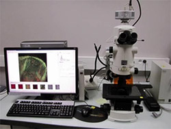
Video microscope DMi8 (Leica)
This fully motorized inverted microscope with automatic focus holding system, epifluorescence and motorized DIC allows the acquisition of images on any sample fixed or not, marked or not by fluorophores. Equipped with a motorized stage and a CO2-regulated thermostatic chamber, this microscope is particularly suitable for automated multicolor kinetics of single or multiple fields. The optical equipment includes an Metal-halide illumination source, DAPI, GFP and TRITC-CY5 fluorescence filters and a set of lenses to meet the majority of video microscopy applications: • Dry lenses: 5x, 10x, 20x (DIC) • Oil lenses: 40x (DIC), 63x (DIC) This microscope is equipped with multiple inserts for slides, dishes and multi-well plates.
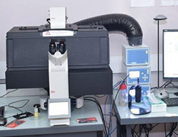
Image processing stations and software
ImageJ & FIJI
Open source software. Their openness, their active community and the richness of their plugins allows the most advanced image processing in many areas. The software architecture integrates many plugins and offers scripting possibilities.
Some applications and their plugins: Co-location: quantification of the colocation of structures on different fluorescent channels (plugins: JACoP, Colocation); Color segmentation: color-based segmentation (plugins: Threshold Colour, Color Segmentation, Color Inspector 3D); Neuron tracing: reconstitution of neuronal morphology (plugins: NeuronJ, NeuriteTracer); Group several stacks together: useful mainly in imaging living cells (plugin: concatenate)
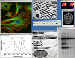
Imaris 9.9
Imaris is proprietary software, owned by the Bitplane company. Specialized in 3D / 4D display, multichannel, it allows visualizing, reconstructing in 3 D volume and surface biological images. It also allows the quantitative analysis of 3D images.
Volocity
Proprietary software, owned by Perkin Elmer. Developed specifically for research in life sciences, the software allows you to visualize, explore and above all quantitatively analyze multi-channel 2D and 3D/4D images. It also includes a deconvolution module suitable for confocal and full-field microscopes. It makes it easy to work in batches.
Huygens
Huygens is proprietary software, published by SVI, dedicated to deconvolution. Deconvolution makes it possible to deflect and defer images from the optical and physical characteristics of the microscope and samples.
ARIVIS Vision4D
Vision4D proprietary software, published by Arivis, which allows the opening of large files. It is a viewer that allows you to work with 2D, 3D-4D, multi-channel images of virtually unlimited sizes and this regardless of the RAM of the workstation. The WebView function makes it easy to share images via the intranet from a web browser.
ICY
ICY is royalty-free software for the processing and analysis of 2D, 3D-4D, multichannel images, which natively integrates ImageJ and is based on a web platform of extensions. It has a large number of plugins/extensions that allow very sharp analyses. In addition, it benefits from an intuitive graphical interface that facilitates the possibilities to realize macros to automatize the analysis and image processing.
