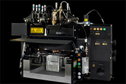
Cytometry & Mass imaging - Equipments
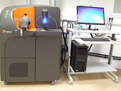
Mass cytometer HELIOS (Fluidigm)
As a complementary tool to flow cytometers, the mass cytometer considerably increases the number of parameters that can be analyzed (>40), while eliminating the problems of overlapping fluorescence spectrums observed with conventional cytometry.
This technology uses non-radioactive metal isotopes of the lanthanide family for sample labeling. These isotopes are absent in biological systems and allow to considerably reduce the background level observed in fluorescence, in particular linked to cellular auto-fluorescence.
Mass cytometry improves the identification of rare cells. It combines the in-depth study of surface markers with intracellular signaling pathways.
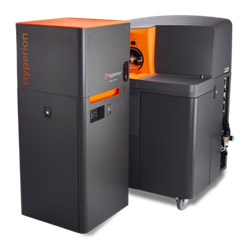
Mass Cytometry Imaging System: HYPERION (Fluidigm)
Combining mass cytometry and imaging capability, the HyperionTM system allows the simultaneous visualization of 4 to 37 markers (complex cellular phenotypes and functional states) on a single histological section, at subcellular resolution in the spatial context of the tissue microenvironment.
Analyzable samples are fresh, frozen or fixed tissue sections, as well as cytological preparations such as cell smears, labeled with antibodies coupled to heavy metal isotopes. Imaging is performed by depletion of tissue areas of very small surface (1mm²)
This technology is particularly relevant in cases where tissues are relatively rare or valuable, such as patient samples.
All raw images need to be processed using algorithms sometimes involving artificial intelligence.
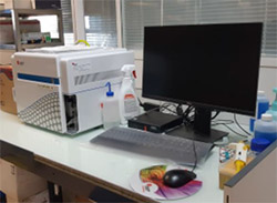
Flow Cytometer CYTOFLEX (Beckman Coulter)
Flow analyzer equipped with 4 lasers emitting respectively at 405 nm, 488 nm, 561 nm and 640 nm. Simultaneous analysis of 15 parameters including 13 fluorescence and 2 morphology (relative size and structure). Automatic calculation of fluorescence compensation during gain changes during acquisition. Counting of cell concentrations in absolute value. The instrument is equipped with an automatic 96-well plate changer. Control of the instrument via the CytExpert software on PC station.
Flow Cytometer LSR FORTESSA II- UV (BD Biosciences)
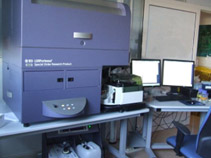
Flow analyzer equipped with 5 lasers emitting respectively at 355, 405, 488, 561 and 640 nm. 20 parameters are analyzed simultaneously, including 18 fluorescence parameters, 2 morphology parameters (relative size and granulosity) and the pulse width for the distinction of aggregates. Fluorescence compensation during and after acquisition. The instrument is equipped with an automatic 96 well plate changer. Control of the instrument via FACSDiVa™ software, on PC station.
Spectral Flow Cytometry AURORA (Cytek)
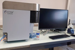
Spectral flow analyzer equipped with 5 lasers emitting respectively at 355nm, 405nm, 488nm, 561nm and 640nm.the instrument has 64 detection channels (APD) for fluorescence and 3 channels for morphological parameters: (FSC and SSC on 488nm laser, and SSC on 405nm laser). It allows the analysis of samples labeled with fluorochomes emitting between 365 and 829nm. Control of the instrument via the SpectroFlo software on PC station.
Cell sorter ARIA-FUSION - UV (BD Biosciences) - 2 identical instruments
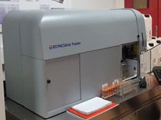
Cell sorter equipped with 5 lasers emitting respectively at 355, 405, 488, 561 and 640 nm: allows the excitation of most fluorochromes in the wavelength range from UV to far red. Simultaneous analysis of 20 parameters, including 18 fluorescence parameters, 2 morphology parameters (size and granulosity) and the pulse width for the distinction of aggregates. Number of sorting lanes: 4 lanes in 1.5 or 5 mL tubes - 2 lanes in 15mL tubes - 1 lane in 6, 24, 48, 96 or 384 well plates. 3 sorting modes: Purity, Yield or Single cell (+ index sorting) Thermostatization of the sample to be sorted 4°, 22°, 37° and 42°C and of the sorted fractions from -4°C to +42°C. Available nozzle diameters are 70μm, 85μm, and 100μm. Data acquisition is performed via the DIVA software.
Cell sorter INFLUX- UV 5Lasers (BD Biosciences)
Cell sorter equipped with 5 lasers emitting respectively at 355, 405, 488, 561 and 640 nm: allows the excitation of most fluorochromes (from UV to far red).
Equipped with a 6-way sorting system, it also performs cloning on multi-well plates or on slides. Simultaneous analysis of 21 parameters, including 19 fluorescence parameters, 2 morphology parameters (size and granulosity) and the pulse width for the distinction of aggregates. Available nozzle diameters are 70μm, 86µm, 100μm and 140μm. Data acquisition is performed via the SORTWARE™ software.
