Cytometry & Mass imaging - Activities
Training and coaching
- Flow, spectral and mass cytometry
- Cell sorting and single-cell isolation
- Mass imaging for molecular, cellular and functional spatial mapping of cells and tissues
- Bioinformatics adapted to the unsupervised analysis of cytometry and imaging data.
Design of experimental procedures
Scientific animations
Technological developments and innovation
Novel multiplex approaches in mass and spectral cytometry
- Phenotypic stratification of leukemic stem cells (LSC) at the origin of relapse in acute myeloid leukemia with transition to an antibody panel adapted to the detection and monitoring of LSC in routine practice using flow cytometry.
- Establishment of functional maps of cancer cells, immune cells and microenvironment interactions on patient tumors, to evaluate the specificity and efficacy of anticancer radio- and chemotherapies.
Bioinformatics: Development of novel analysis strategies
- Unsupervised analysis of flow, spectral and mass cytometry data
- Quantitative analysis of mass imaging data

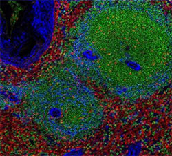
Multiplex imaging of a human spleen sample by mass cytometry

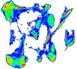
Characterization of leukemic stem cells in acute myeloid leukemia in a cohort of 45 patients
Catégorie de la page:
